A spasticity assessment often ends with a single number from a scale. However, the resistance you feel during passive movement has several sources, such as spasticity, spastic dystonia and contracture [1,2]. A single number cannot tell them apart, so it cannot tell you what to treat.
This guide shows how to assess spasticity in the order you would do it in the clinic. First, it covers what to ask and what to look at. Then it shows how to test active movement and how to stretch slowly and quickly. Finally, it shows how to turn the findings into goals you can retest.
What a spasticity assessment should answer
To begin with, spasticity is only one form of muscle overactivity after an upper motor neuron lesion. Yelnik and colleagues describe three forms [2]. Each one shows up under different conditions:
- Spasticity is an increased, velocity-dependent response to stretch. You detect and measure it at rest.
- Spastic dystonia is tonic muscle activity at rest, with no primary trigger. It deforms joints and postures.
- Spastic co-contraction is excessive activity in the antagonist during a voluntary command on the agonist. You detect it during voluntary effort.
All three are stretch-sensitive [2,3]. We describe them, together with the other signs, in Upper Motor Neuron Syndrome: Positive and Negative Signs.
Before any treatment, Yelnik and colleagues ask three questions [2]. First, is the muscle overactivity a problem, and in what way? Second, is it the main cause of the disability, or only one of several causes? Third, is it limited to one muscle group, or is it widespread? A good spasticity assessment answers all three.
| Step | What you do | What it tells you |
|---|---|---|
| 1 | Take a history | The patient’s problem and what makes the tone worse |
| 2 | Standardize the conditions | A result you can compare at the next visit |
| 3 | Observe the limb at rest | Spastic dystonia and resting posture |
| 4 | Test active movement | What the patient can do alone, co-contraction and repeatability |
| 5 | Stretch slowly | Passive range of motion and contracture |
| 6 | Stretch fast | Angle of catch or clonus and the spasticity grade |
| 7 | Assess function | The impact on daily life |
| 8 | Set goals and retest | What success means for this patient |
Step 1: Take a history and look for triggers
First of all, ask about the patient’s own problem. That includes pain, discomfort, spasms and clonus, as well as washing, dressing, sitting and walking [2,4]. For patients with cognitive impairment, reports from caregivers are particularly important [4].
Then look for anything that makes the tone worse. Dromerick lists pain, fatigue, anxiety, wounds, fractures and systemic infection, among other factors [4]. His list also includes ingrown toenails, heterotopic ossification, urinary retention and constipation. Yelnik and colleagues add that such painful triggers must be treated before the muscle overactivity itself. In fact, the patient may not notice them if sensation is reduced [2].
Finally, ask what the overactivity does for the patient, because not every patient with spasticity needs treatment [2]. For example, some patients with severe paresis stand with the help of quadriceps overactivity [2]. Similarly, elbow flexor overactivity may let them carry a bag.
Step 2: Standardize the conditions
A spasticity assessment is only useful if you can repeat it. Yet muscle overactivity is not constant. For example, the time of day and the position of other joints, including the neck, can change it [2]. Similarly, stress, outside temperature and any painful stimulus can change it. Therefore, test at a similar time of day and in the same body position at every visit [2,4]. Also note both in your records.
In our practice, we start with the patient sitting, where the trunk has to work against gravity. Then we move to supine, where the patient no longer has to control the trunk against gravity. Whichever positions you choose, use the same ones at every visit.
Step 3: Observe the limb at rest
Before you move anything, look. Spastic dystonia is the form you can recognize most easily just by looking, because it deforms joints and posture [2]. In the arm, for instance, the shoulder may stay adducted and internally rotated. Meanwhile, the elbow, wrist and fingers stay flexed, and the forearm stays pronated [2]. In the leg, spastic dystonia often involves the plantar flexors and causes an equinovarus foot [2]. When it involves the toe flexors, it causes toe clawing.
Also watch the patient sit. Clonus can appear while the patient simply tries to sit relaxed [2]. As a result, it can be hard to keep the feet on the footplates of a wheelchair.
Step 4: Start with active movement
In our practice, we start with active movements. They give us a reference for what the patient can do alone, before we move the limb ourselves.
Ask the patient to move actively, as far as possible, against the muscle group you are assessing [2]. The movement stops where the agonist can no longer overcome passive resistance and spastic co-contraction in the antagonist. Thus, this angle is the active range of motion [2,3].
After that, ask for rapid alternating movements over the same range [2]. The patient does as many as possible in a set time, for example 15 seconds. Gracies and colleagues use this step to check how repeatable the active movement is [3]. Moreover, in the experience of Yelnik and colleagues, it reveals spastic dystonia or co-contraction in a simple examination [2].
While the patient works, watch the rest of the body as well. Unwanted activity in distant muscles during effort, such as associated reactions, is another form of overactivity [2].
Step 5: Stretch slowly to find the passive range
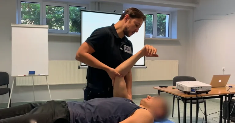
Now move each muscle group yourself, very slowly, below the speed that triggers a stretch reflex. Yelnik and colleagues call this slow velocity V1 [2]. The angle at which the soft tissues offer maximum resistance is the passive range of motion [2]. This angle reflects the length and extensibility of the soft tissues [3]. In other words, it is your measure of contracture. Then record it in degrees.
However, severe spastic dystonia can look like a fixed contracture. To tell them apart, a physician sometimes performs a temporary nerve or motor point block with a local anesthetic [2].
Step 6: Stretch fast to find the catch
Next, stretch the same muscle group as fast as you can. This is velocity V3 in the Tardieu method [1,2]. At this point, two findings matter. First, the angle at which a catch or clonus appears shows the threshold of the stretch reflex. Second, the type of reaction at that angle gives the grade [2].
Thibaut and colleagues summarize the grades of the Modified Tardieu Scale [1]:
| Grade | Muscle reaction |
|---|---|
| 0 | No resistance throughout the passive movement |
| 1 | Slight resistance throughout the passive movement, no clear catch at a precise angle |
| 2 | Clear catch at a precise angle, interrupting the movement, followed by release |
| 3 | Fatigable clonus lasting less than 10 seconds while you maintain the pressure |
| 4 | Unfatigable clonus lasting more than 10 seconds while you maintain the pressure |
| 5 | Joint is fixed |
The scale also defines three velocities [1]. V1 is as slow as possible, V2 is the speed of the limb falling under gravity, and V3 is as fast as possible.
Why compare the slow and the fast stretch
Comparing the two stretches is what makes this step useful. Patrick and Ada examined the elbow flexors and ankle plantar flexors of 16 people three years after stroke [5]. In both muscle groups, the Tardieu Scale matched a laboratory measure of spasticity in 100% of cases. By contrast, the Ashworth Scale agreed in 63%. Moreover, whenever the Ashworth Scale overestimated spasticity, the participant had a contracture.
If clonus appears, time it while you keep the pressure on. We explain the full test in Ankle Clonus: Causes, Testing and What It Means.
Clinical tip
For each muscle group, record three angles in the same position. These are the active angle, the slow passive angle and the angle of catch. Gracies and colleagues measure the same three angles in the first three steps of their Five Step Assessment [3]. As a result, you can see at a glance whether contracture, spasticity or poor active control limits the joint.
Step 7: Link the findings to function
A finding in a spasticity assessment matters only if it affects the patient’s life. The right tools depend on how much motor control the patient has [2].
Severe paresis. Here the problems are mostly passive: hygiene, dressing, limb position, pain, sitting and the risk of pressure sores [2]. The Disability Assessment Scale covers four of these domains, namely hygiene, dressing, limb position and pain [1,2]. Likewise, the patient’s disability scale and the carer burden rating scale cover tasks such as cleaning the palm [2].
Fairly good motor control. Here the question is whether overactivity limits active movement. For the leg, use walking tests over 10 meters, 2 minutes or 6 minutes [2]. For the arm, Yelnik and colleagues point to the Modified Frenchay Scale, which rates 10 real-life tasks [2]. On the other hand, general scales of daily living such as the Barthel Index are less useful here [2]. That is because patients can complete many daily tasks without using the affected arm.
Where the Modified Ashworth Scale fits in a spasticity assessment
Of course, the Modified Ashworth Scale is quick and easy to use [1]. Bohannon and Smith described it in 1987 [6]. They graded the elbow flexors of 30 patients and agreed on 86.7% of their ratings.
Its limits, however, are well documented. The scale measures resistance to passive movement, not spasticity [7]. Pandyan and colleagues even treat it as a nominal measure until the ambiguity between grades 1 and 1+ is resolved [7]. In addition, it scores a mix of contracture, spastic dystonia and spasticity [1,2]. Fleuren and colleagues tested the original Ashworth Scale in 30 patients [8]. The intraclass correlation between raters was 0.58 for the elbow flexors and 0.63 for the knee extensors. Furthermore, the rater strongly influenced the score. Consequently, they concluded that its validity and reliability are insufficient to measure spasticity.
So record the Modified Ashworth Scale if your setting requires it. Just do not let it stand in for the whole spasticity assessment. The Tardieu method fits Lance’s definition more closely, although its validity and reliability still need more research [9]. We discuss the definition in Lower Limb Spasticity: 5 Things Physiotherapists Should Know.
Step 8: Set goals and retest
Finally, turn the findings into goals. Yelnik and colleagues recommend a list of personal objectives that you can measure separately, which then drives treatment [2]. An international consensus also prefers an individual approach to treatment and outcome measurement [10]. Goal attainment scaling is one way to do this [11]. With this method, each goal is rated on a 5-point scale from −2 to +2.
Dromerick suggests objective or semi-objective endpoints [4]. Examples include range of motion, spasm frequency, pain scales, the fit of a brace or splint, and the Ashworth Scale. Retest them in the same position, and then compare the results with the goals you set at the start. That closes the loop of a spasticity assessment.
Key points
- A spasticity assessment separates spasticity, spastic dystonia and spastic co-contraction from contracture and weakness.
- Ask about the patient’s problems and look for triggers such as pain, infection and constipation before you test.
- Test in the same position and at a similar time of day at every visit.
- Start with active movement, then stretch slowly and fast. For each muscle group, record the active angle, the slow passive angle and the angle of catch.
- Link every finding to passive or active function.
- The Modified Ashworth Scale records resistance to passive movement, not spasticity.
- Set measurable personal goals and retest them in the same conditions.
References
- Thibaut A, Chatelle C, Ziegler E, Bruno MA, Laureys S, Gosseries O. Spasticity after stroke: physiology, assessment and treatment. Brain Inj. 2013;27(10):1093-1105. doi:10.3109/02699052.2013.804202
- Yelnik AP, Simon O, Parratte B, Gracies JM. How to clinically assess and treat muscle overactivity in spastic paresis. J Rehabil Med. 2010;42(9):801-807. doi:10.2340/16501977-0613
- Gracies JM, Bayle N, Vinti M, et al. Five-step clinical assessment in spastic paresis. Eur J Phys Rehabil Med. 2010;46(3):411-421. PMID: 20927007
- Dromerick AW. Clinical features of spasticity and principles of treatment. In: Gelber DA, Jeffery DR, eds. Clinical Evaluation and Management of Spasticity. Humana Press; 2002:13-26. doi:10.1007/978-1-59259-092-6_2
- Patrick E, Ada L. The Tardieu Scale differentiates contracture from spasticity whereas the Ashworth Scale is confounded by it. Clin Rehabil. 2006;20(2):173-182. doi:10.1191/0269215506cr922oa
- Bohannon RW, Smith MB. Interrater reliability of a modified Ashworth scale of muscle spasticity. Phys Ther. 1987;67(2):206-207. doi:10.1093/ptj/67.2.206
- Pandyan AD, Johnson GR, Price CI, Curless RH, Barnes MP, Rodgers H. A review of the properties and limitations of the Ashworth and modified Ashworth Scales as measures of spasticity. Clin Rehabil. 1999;13(5):373-383. doi:10.1191/026921599677595404
- Fleuren JF, Voerman GE, Erren-Wolters CV, et al. Stop using the Ashworth Scale for the assessment of spasticity. J Neurol Neurosurg Psychiatry. 2010;81(1):46-52. doi:10.1136/jnnp.2009.177071
- Haugh AB, Pandyan AD, Johnson GR. A systematic review of the Tardieu Scale for the measurement of spasticity. Disabil Rehabil. 2006;28(15):899-907. doi:10.1080/09638280500404305
- Sheean G, Lannin NA, Turner-Stokes L, Rawicki B, Snow BJ. Botulinum toxin assessment, intervention and after-care for upper limb hypertonicity in adults: international consensus statement. Eur J Neurol. 2010;17(suppl 2):74-93. doi:10.1111/j.1468-1331.2010.03129.x
- Turner-Stokes L. Goal attainment scaling (GAS) in rehabilitation: a practical guide. Clin Rehabil. 2009;23(4):362-370. doi:10.1177/0269215508101742