Ankle clonus is one of the fastest signs to check in a neurological exam. You dorsiflex the foot briskly, keep the pressure on, and the calf answers with a series of rhythmic beats. The beats tell you that the stretch reflex is hyperexcitable. However, they do not tell you how severe the spasticity is, or what to treat first.
This guide explains what ankle clonus is, how to perform and grade a clonus test, and what causes clonus. Finally, it covers what the finding means for your patient and what the evidence says about managing it.
What is ankle clonus?
Clonus is an involuntary, rhythmic muscle contraction that follows a sudden muscle stretch [1]. It results from a lesion of the descending motor pathways and often comes with spasticity and increased tendon reflexes [1]. Boyraz and colleagues list the ankle, patella, triceps surae, wrist, jaw and biceps brachii among the sites where it appears [2]. The ankle is the most common one [3].
The rhythm is remarkably regular. Clonus usually beats at 5 to 8 Hz, and one cycle of ankle clonus lasts about 160 to 200 ms [2]. Plantar flexion takes about 45% of each cycle and dorsiflexion about 55%. The first beat is always the longest. The following beats get shorter and settle by the fourth or fifth cycle [2].
Clonus is one of the positive signs of the upper motor neuron syndrome, together with spasticity, increased tendon reflexes, the Babinski sign and spasms [4]. In fact, Trompetto and colleagues trace spasticity, clonus and increased tendon reflexes to one cause: hyperexcitability of the stretch reflex [4]. We describe the whole syndrome in Upper Motor Neuron Syndrome: Positive and Negative Signs.
Clonus and spasticity are related, but not the same
The two signs share a reflex, yet they do not always move together. Li and Francisco review older reports of cordotomy, a surgical cut in the spinal cord [5]. Extensive cuts that damaged the medial reticulospinal tract reduced spasticity drastically. However, tendon hyperreflexia, clonus and adductor spasms persisted.
Likewise, clinical scales do not always agree. In 85 people with spinal cord injury, Priebe and colleagues found that common spasticity scales correlated poorly with each other [6]. For ankle clonus, the strongest link was with the Achilles tendon reflex. The authors therefore concluded that each scale captures a different aspect of spasticity.
So record clonus as a finding of its own. One score does not predict the other, so a tone scale such as the Modified Ashworth Scale cannot replace the clonus test. For how spasticity, contracture and clonus differ, see Lower Limb Spasticity: 5 Things Physiotherapists Should Know.
How to do a clonus test at the ankle
The clonus test uses the stimulus that defines clonus: a sudden stretch of the calf, held in place [1]. The Modified Tardieu Scale grades clonus while the examiner maintains the pressure after the stretch [7].
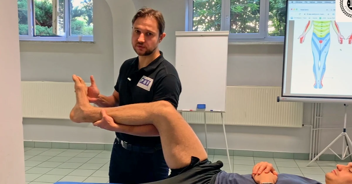
- Position the patient. In our practice, we test with the patient supine and the leg flexed at the hip and knee. This position lets the whole body relax without building up tension before the test.
- Use the same position every time. Rossi and colleagues showed that clonus duration depends on the joint angle, and probably on how far the triceps surae is stretched [8]. So repeat it at every visit and write it down.
- Dorsiflex the foot briskly. Clonus is a response to a sudden stretch [1].
- Keep the pressure. Hold the foot in dorsiflexion while you count the beats or time them with a stopwatch.
- Release and observe. Clonus usually depends on position, so a change in position usually stops it [3].
- Test both sides. Then record the result separately from muscle tone [6].
Clinical tip
Stretch is not the only trigger. Dimitrijevic and colleagues showed that other inputs, such as skin stimulation, can start clonus [9]. Many patients also report more tone, spasms and clonus when they are tired or anxious [3]. Keep your grip, the position and the setting the same each time, and note anything that may have changed the result.
Of course, the clonus test is only one part of a full exam. We cover the rest in Spasticity Assessment: A Step-by-Step Clinical Exam.
How to grade ankle clonus
For example, Boyraz and colleagues used a 7-point reflex scale that separates unsustained from sustained clonus [1].
| Grade | Finding |
|---|---|
| 0 | None |
| 1 | Hyporeflexia |
| 2 | Normal |
| 3 | Pathologically brisk, no clonus |
| 4 | Unsustained clonus, fewer than 4 beats |
| 5 | Unsustained clonus, more than 4 beats |
| 6 | Sustained clonus |
In contrast, the Modified Tardieu Scale uses time instead of beats [7]. Grade 3 is fatigable clonus that lasts less than 10 seconds while you maintain the pressure. Grade 4 is unfatigable clonus that lasts more than 10 seconds.
How to measure change over time
Grades work well for documentation. To show change, though, measure duration. Manella and colleagues tested 40 people with chronic spinal cord injury, using a standardized drop test to provoke ankle clonus [10]. Clonus duration timed with a stopwatch was highly reliable within and between raters, and over one week.
The number of oscillations in the first 10 seconds was also reliable. In addition, it correlated strongly with a clinical clonus score (r = 0.86) and with the soleus H-reflex (r = 0.77) [10]. Counting oscillations required motion capture, while a stopwatch needs nothing more than a watch. So time the clonus in the same position before and after treatment.
What causes clonus?
The exact mechanism is still unclear, and two explanations compete [2]. Each, though, has experimental support.
Self-excitation of the stretch reflex
The most widely accepted explanation is self-excitation of a hyperactive stretch reflex [2]. The stretch fires the reflex and the calf contracts. Then, as the muscle relaxes, your pressure stretches it again and fires the next beat. Boyraz and colleagues describe clonus as sustained by stretch reflexes during muscle relaxation [1].
Hidler and Rymer modeled this loop at the ankle [11]. Oscillations appeared when two conditions met: a long reflex delay, typical of distal leg muscles, and increased motoneuron excitability. Specifically, the key change was a lower motoneuron firing threshold, not a higher reflex gain.
Experiments in patients also point the same way. In 4 patients, the same authors found that clonus frequency changed with the applied load [12]. Overall, clonus behaved like a stable cycle driven by peripheral receptors. When Rossi and colleagues blocked group I afferents from the calf by compressing the upper third of the lower leg, ankle clonus disappeared within 10 minutes [8].
A central generator in the spinal cord
The alternative explanation is a rhythm generator in the spinal cord [2]. Peripheral events switch it on, and then it drives the motoneurons at its own rate. Walsh applied a rhythmic force to the foot in nine patients with sustained clonus [13]. The clonus did not lock to the external rhythm, so he concluded that a spinal generator sets its rate.
Dimitrijevic and colleagues could not change the rate of clonus either [9]. In people with spinal cord injury, Beres-Jones and colleagues recorded synchronous clonic bursts in the soleus, gastrocnemius and tibialis anterior [14]. The bursts were not consistently linked to muscle stretch. They looked alike during clonus, stepping and standing.
What the debate means in the clinic
Neither view explains everything. Beres-Jones and colleagues concluded that central mechanisms and peripheral events probably interact [14]. Boyraz and colleagues also suggest that group II afferents may contribute alongside group Ia afferents [2].
Still, for your exam the message is simple. The stretch you apply matters, so standardize it. Other inputs matter too, so control what you can: skin contact, position, fatigue and anxiety [3,9].
What ankle clonus means for your patient
Function
Clonus can be a mere curiosity or a real disability [3]. Dromerick notes that it can prevent functional use of a limb, make splinting difficult, and distract or embarrass the patient. In addition, severe clonus can limit mobility and cause fatigue, and some patients lose the ability to stand or walk [1].
Ask the patient when the shaking starts and what stops it, and watch functional tasks. Clonus usually appears with the muscle on stretch [3]. Because position can stop it, the answer often points to a practical solution in daily tasks.
A sign that needs an explanation
Clonus is an upper motor neuron sign [15]. After a stroke or spinal cord injury, it fits the picture. In a patient referred for something else, however, it needs a medical explanation.
For example, Mirza and colleagues describe a woman with cauda equina syndrome and lower limb clonus [15]. Clonus and her history raised the possibility of a second diagnosis, so the team also imaged the brain and the cervical and thoracic spinal cord. In the end, the clonus was attributed to the cauda equina compression and later resolved after surgery. That is why, if you find ankle clonus that no known diagnosis explains, you should report it to the referring physician.
Managing ankle clonus
Current treatments for spasticity act mainly on its positive signs, clonus included [3]. However, evidence specific to clonus is thin, and most studies are small.
Cold
Cold is a treatment physiotherapists can apply themselves. Boyraz and colleagues treated 19 patients with sustained ankle clonus [1]. The legs were immersed in a 15°C whirlpool for 20 minutes, once a day, for 10 sessions. Afterwards, clinical ankle clonus was gone in 16 of the 19 patients. However, it came back after an average of 50 hours, with a range of 1 to 7 days.
The effect outlasted the cooling of the leg, and the authors suggest a change at the spinal level [1]. In contrast, a group that received rehabilitation alone for about 3 weeks showed no significant change. Also note that patients with peripheral vascular disease or cold intolerance were not assigned to cold. The groups were small and not randomized, so treat this as a promising signal, not proof.
Medication and injections
Medication is the physician’s decision, but the data are worth knowing. In the same study, tizanidine at the highest tolerated dose lowered Ashworth scores but had no significant effect on ankle clonus [1]. The authors cite several studies in which centrally acting antispastic drugs did not change clonus significantly, and some in which baclofen had a stronger effect.
A systematic review of botulinum toxin type A included 14 studies with 181 patients [16]. All of them showed improvement in clonus, but the improvement was statistically significant in only 6. The authors called the evidence preliminary, because the clonus measures varied and lacked proven validity and reliability. Phenol injections are another option [2].
Active training
Training data come from people with motor-incomplete spinal cord injury. Manella and Field-Fote studied 18 people before and after 12 weeks of body-weight-supported locomotor training [17]. Ankle clonus decreased, but the change did not reach statistical significance (p = 0.09). The authors read the overall pattern as a reduction in extensor spasticity.
Key points
- Ankle clonus is an involuntary, rhythmic contraction of the calf after a sudden stretch. It signals a hyperexcitable stretch reflex after an upper motor neuron lesion.
- Test it the same way every time: same position, a brisk dorsiflexion, then hold the pressure.
- Grade it by beats (unsustained or sustained) or by time (Modified Tardieu Scale grade 3 under 10 seconds, grade 4 over 10 seconds). For follow-up, time it with a stopwatch.
- Clonus does not measure the severity of spasticity, so record it separately from muscle tone.
- Clonus that no known diagnosis explains needs a medical explanation.
- Cold, botulinum toxin and some medications can reduce clonus, but the evidence is limited.
References
- Boyraz I, Oktay F, Celik C, Akyuz M, Uysal H. Effect of cold application and tizanidine on clonus: clinical and electrophysiological assessment. J Spinal Cord Med. 2009;32(2):132-139. doi:10.1080/10790268.2009.11760764
- Boyraz I, Uysal H, Koc B, Sarman H. Clonus: definition, mechanism, treatment. Med Glas (Zenica). 2015;12(1):19-26. PMID: 25669332
- Dromerick AW. Clinical features of spasticity and principles of treatment. In: Gelber DA, Jeffery DR, eds. Clinical Evaluation and Management of Spasticity. Humana Press; 2002:13-26. doi:10.1007/978-1-59259-092-6_2
- Trompetto C, Marinelli L, Mori L, et al. Pathophysiology of spasticity: implications for neurorehabilitation. Biomed Res Int. 2014;2014:354906. doi:10.1155/2014/354906
- Li S, Francisco GE. New insights into the pathophysiology of post-stroke spasticity. Front Hum Neurosci. 2015;9:192. doi:10.3389/fnhum.2015.00192
- Priebe MM, Sherwood AM, Thornby JI, Kharas NF, Markowski J. Clinical assessment of spasticity in spinal cord injury: a multidimensional problem. Arch Phys Med Rehabil. 1996;77(7):713-716. doi:10.1016/s0003-9993(96)90014-3
- Thibaut A, Chatelle C, Ziegler E, Bruno MA, Laureys S, Gosseries O. Spasticity after stroke: physiology, assessment and treatment. Brain Inj. 2013;27(10):1093-1105. doi:10.3109/02699052.2013.804202
- Rossi A, Mazzocchio R, Scarpini C. Clonus in man: a rhythmic oscillation maintained by a reflex mechanism. Electroencephalogr Clin Neurophysiol. 1990;75(2):56-63. doi:10.1016/0013-4694(90)90152-a
- Dimitrijevic MR, Nathan PW, Sherwood AM. Clonus: the role of central mechanisms. J Neurol Neurosurg Psychiatry. 1980;43(4):321-332. doi:10.1136/jnnp.43.4.321
- Manella KJ, Roach KE, Field-Fote EC. Temporal indices of ankle clonus and relationship to electrophysiologic and clinical measures in persons with spinal cord injury. J Neurol Phys Ther. 2017;41(4):229-238. doi:10.1097/NPT.0000000000000197
- Hidler JM, Rymer WZ. A simulation study of reflex instability in spasticity: origins of clonus. IEEE Trans Rehabil Eng. 1999;7(3):327-340. doi:10.1109/86.788469
- Hidler JM, Rymer WZ. Limit cycle behavior in spasticity: analysis and evaluation. IEEE Trans Biomed Eng. 2000;47(12):1565-1575. doi:10.1109/10.887937
- Walsh EG. Clonus: beats provoked by the application of a rhythmic force. J Neurol Neurosurg Psychiatry. 1976;39(3):266-274. doi:10.1136/jnnp.39.3.266
- Beres-Jones JA, Johnson TD, Harkema SJ. Clonus after human spinal cord injury cannot be attributed solely to recurrent muscle-tendon stretch. Exp Brain Res. 2003;149(2):222-236. doi:10.1007/s00221-002-1349-5
- Mirza AB, Akhbari M, Lavrador JP, Maratos EC. Atypical cauda equina syndrome with lower limb clonus: a literature review and case report. World Neurosurg. 2020;134:507-509. doi:10.1016/j.wneu.2019.10.198
- Thanikachalam V, Phadke CP, Ismail F, Boulias C. Effect of botulinum toxin on clonus: a systematic review. Arch Phys Med Rehabil. 2017;98(2):381-390. doi:10.1016/j.apmr.2016.09.126
- Manella KJ, Field-Fote EC. Modulatory effects of locomotor training on extensor spasticity in individuals with motor-incomplete spinal cord injury. Restor Neurol Neurosci. 2013;31(5):633-646. doi:10.3233/RNN-120255